Our Hours: Monday - Friday: 8:00 AM - 5:00 PM EST
Menu
Contact Us

Diagnostic Imaging

Why is diagnostic imaging (MRI, CT Scan, X-Ray) often needed when diagnosing neck and back pain related issues?

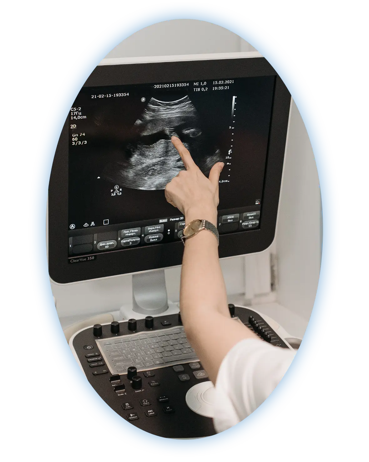
When you consult with a physician regarding a back pain diagnosis, he or she will typically request one or more imaging scans. While a physical examination can help your physician gain a better understanding of your symptoms, imaging scans will likely be needed to confirm the specific cause of your discomfort.

What types of diagnostic imaging are used on the spine?

Based on the type of back pain diagnosis your physician suspects, you may be scheduled for any of the following imaging scans:

MRI Scans are integral to ensuring accurate diagnosis in many cases
  • X-ray. Depending on the part of your spine that is being examined, you’ll be asked to either lie on your back on an X-ray table or stand in a comfortable position while an X-ray beam is focused on a specific area of your body. One or more images will then be taken and processed, allowing your physician to view the composition of your spinal bones in detail.
  • CT (computed tomography) scan. A CT scan is a more detailed type of X-ray that produces cross-sectional images. During this test, you’ll be asked to lie on a table as a scanning machine circles your body. The resulting images will be sent to a computer monitor and processed by a technician, then provided to your physician to be evaluated for your back pain diagnosis.
  • Bone scan. In this procedure, a small amount of radioactive material known as a tracer will be injected into a vein. The tracer will then circulate throughout your body so that it can be absorbed by your bones. During the scan, you’ll be asked to lie on a table while a small electronic arm passes a camera over your body. In the resulting images, any bone abnormalities will appear lighter or darker in contrast to the surrounding healthy bone tissue.
  • MRI (magnetic resonance imaging) scan. For a spinal MRI, you’ll be asked to lie on a table, where a scanner will produce radio waves and capture their path as they bounce off your body. This information will then be used to produce pictures of your spinal anatomy that will be provided to your physician to be evaluated for your back pain diagnosis. While X-rays and CT scans can only produce images of your spinal bones, MRIs can also show the spaces in between the bones that the nerves pass through.

Once you’ve received an official diagnosis of the cause of your back pain, you’ll be able to get started with treatment. Your imaging scans may be referenced at various points during the treatment process.

Schedule Your Appointment

Our Patients Stories

Free MRI Review

Looking for a second opinion?
Start Your Free MRI Review

Melbourne: 709 S Harbor City Blvd, Ste 110, Melbourne, FL 32901
Orlando: 9145 Narcoossee Rd, Ste A200, Orlando FL 32827
Titusville: 494 N Washington Ave, Titusville, FL 32796

Copyright © Total Spine and Orthopedics - Icons From FlatIcon

crossmenu